Anisotropic dispersion of excitonic bands of the single-crystal pentacene (001) surface as measured by low-energy angle-resolved high-resolution electron energy-loss spectroscopy
Result of the Month
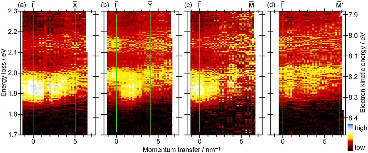
Figure 1: 2D mapping image of angle-resolved HREELS spectra of pentacene single-crystal (001), taken in different directions. The right axis indicates the electron kinetic energy for each spectrum, and the left axis is based on the common energy-loss range for all four images.
Sample damage by electron irradiation and charging of the sample due to insufficient electron conductance are the main challenges to measure molecular solid samples using HREELS (and ARPES). These challenges are overcome by two tricks. One is the reduction of the incident electron flux. The other is illumination of the sample with visible light (from a diode laser) to utilize the photoconduction of the sample for cancelling the sample charging. Anisotropic dispersion was observed thanks to those solutions.
Due to low electron flux during the measurement to avoid sample damage, it took approximately 24 hours to obtain each 2D spectrum. This highlights the advantage of using a hemispherical electron analyser with 2D detector for HREELS measurements, since it would take weeks to measure the same spectra with traditional single channel instruments.
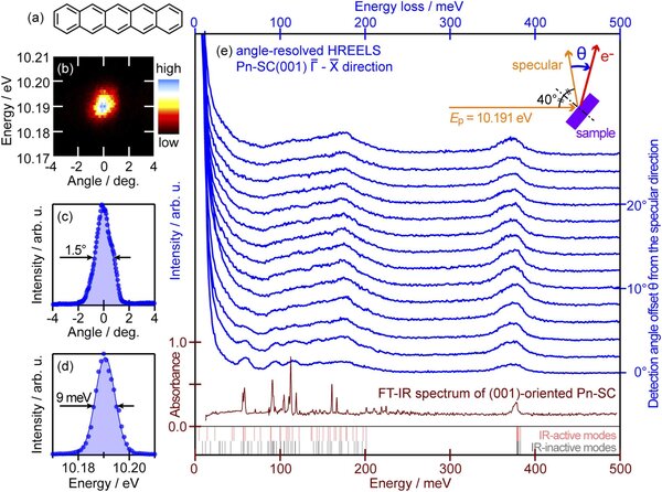
Fighure 2: (a) Molecular structure of pentacene. (b) Typical electron beam spot for the specular reflection from the pentacene single-crystal (001) sample. (c) Profile of (b) in the angular direction. (d) Profile of (b) in the energy direction. (e) angle-resolved HREELS spectra in the Γ̅−X̅ direction. The red curve represents the FT-IR absorbance spectrum from a reference. Results of a quantum chemical calculation for the frequencies of IR-active and IR-inactive vibration modes for a single pentacene molecule are displayed as pale-red and gray vertical bars, respectively. (Inset) Schematic drawing of the measurement geometry.
The specular reflection demonstrates the performance of the instrument (total experimental resolution of 9 meV in the current measurement settings). Excitons are intrinsically much broader, which limits the experimental resolution in the figure 2(e).
The primary energy of the electron source is chosen to be 10 eV based on the following consideration. Low energy is preferred from experience in previous ARPES measurements. This is presumably because the topmost molecular layer of single-crystal pentacene is different from that of the bulk, and the electronic properties of bulk single-crystal pentacene cannot be probed unless the primary electron energy is reduced as low as possible to increase the electron inelastic mean free path to surpass the molecular layer thickness (about 1.4 nm). On the other hand, a primary energy higher than 10 eV is needed to reach the Brillouin zone boundaries using R4000 analyser.