In-situ probing of the Fischer-Tropsch reaction on Co single crystal surfaces up to 1 bar

Result of the Month

Author: Patrick Lömker, David Degerman, Christopher M. Goodwin, Mikhail Shipilin, Peter Amann, Gabriel L. S. Rodrigues, Fernando Garcia-Martinez, Raffael Rameshan, Jörgen Gladh, Hsin-YiWang, Markus Soldemo, Alexander Holm, Steffen Tober, Jan-Christian Schober, Leon Jacobse, Vedran Vonk, Robert Gleißner, Heshmat Noei, Zoltan Hegedues, Andreas Stierle, Christoph Schlueter & Anders Nilsson Institute: ''Stockholm University'' Nature Communications
URL: https://www.nature.com/articles/s41467-025-56082-8
Date: 11/2025
Instruments: XPS Lab, HiPP-2

The surface chemistry of the Fischer-Tropsch catalytic reaction over Co has still several unknows. Here, we report an in-situ X-ray photoelectron spectroscopy study of Co(0001) and Co(10-14), and in-situ high energy surface X-ray diffraction of Co during the Fischer-Tropsch reaction at 0.15 bar - 1 bar and 406 K - 548 K in a H2/CO gas mixture. We find that these Co surfaces remain metallic under all conditions and that the coverage of chemisorbed species ranges from 0.4–1.7 monolayers depending on pressure and temperature. The adsorbates include CO on-top, C/-CxHy and various longer hydrocarbon molecules, indicating a rate-limiting direct CO dissociation pathway and that only hydrocarbon species participate in the chain growth. The accumulation of hydrocarbon species points to the termination step being rate-limiting also. Furthermore, we demonstrate that the intermediate surface species are highly dynamic, appearing and disappearing with time delays after rapid changes in the reactants’ composition. 

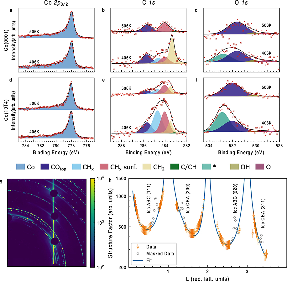
Surface state of Co(0001) sample at indicated temperatures in an atmosphere of CO:H2 1:2 at 1 bar studied by hard X-ray photoelectron spectroscopy (HAXPES) utilizing 4600 eV photons at 0.3° incidence. a shows Co 2p3/2 core-level spectra b displays the C 1 s region and c depicts the O 1 s region. Subplots d, e, f show the same conditions as a, b, c but for a Co(101¯4) surface. The columns of XPS data have constant scaling of the vertical axis. Subplot g shows a representative figure of the high energy surface X-ray diffraction HESXRD data at 67.4 keV of the Co(0001) crystal (full set is shown in SI). The detector is protected by beam stops at the bulk Bragg peak positions. h X-ray structure factor extracted from the 2D diffraction data shown in g at reaction conditions at 496 K and 1 bar reaction mixture (1:2 CO:H2), data from the hcp part of the surface used for the fit (orange circles with vertical lines indicating an estimation 10% relative error), data from the fcc part (grey circles), fit result (solid line).