Sub-Angstrom Noninvasive Imaging of Atomic Arrangement in 2D Hybrid Perovskites
Result of the Month

qPlus-based STM and ncAFM imaging of the RPP surface
The past few years have witnessed a surge of worldwide research interest and rapid growth in the field of two-dimensional (2D) Ruddlesden–Popper halide perovskites (RPPs). Both fundamental interest and technology relevance of 2D RPPs are closely linked to their quantum well structures consisting of insulating organic layers sandwiched between conducting inorganic lead-halide frameworks. The presence of additional soft insulating organic layers in 2D RPPs versus three-dimensional (3D) counterparts introduces the two-dimensionality, abundance of emergent quantum phenomena, and also leads to novel light-matter interaction and significantly enhanced photo- and chemical stability. However, due to insulating nature and softness of organic layers as well as “buried” inorganic framework, accessing real-space atomic arrangement, and understanding the related effects in 2D RPPs remain a grand challenge.
To this end, a NUS research team led by Prof. Jiong LU, used combined low-temperature STM and ncAFM imaging techniques to resolve atomic structures of both inorganic framework and organic layers in the prototypical hybrid lead-halide RPP crystal (Figure A). The STM imaging resolves the reconstruction of the inorganic octahedral framework and unveils the twin-domain composition of the RPPs. On the other hand, ncAFM imaging with a CO-functionalized tip enabled a noninvasive visualization of the cooperative reconstruction of the on-surface cations with sub-angstrom resolution (Figure B,C). The reconstruction of the on-surface organic layers, presented by a well-ordered array of the pairs of butylammonium cations (Figure (D)), was found to be intimately interlocked with the deformation of the inorganic lattice through hydrogen bonding interactions, which is corroborated by density functional theory calculations.
The team also conducted the atomic-scale imaging of the electrostatic potential variation across the twin-domain walls using Kelvin Probe Force Microscopy technique, which reveals the alternating quasi-one-dimensional (1D) electron and hole channels at neighbouring interdomain boundaries, possibly responsible for efficient separation of photoexcited electron-hole pairs and long-distance exciton diffusion in RPPs.
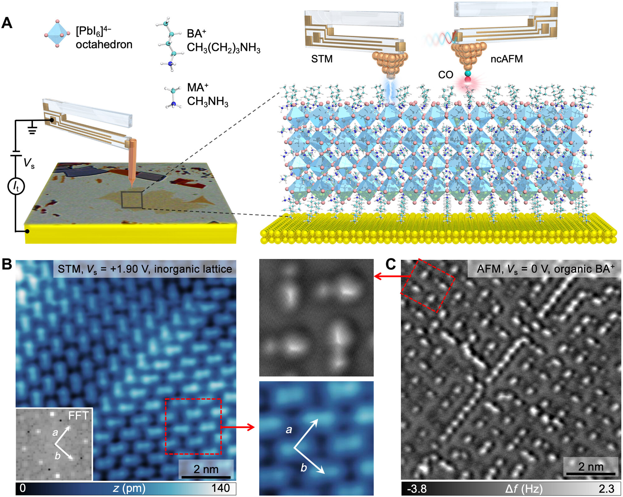
Figure: (A) Schematics showing a combined STM and ncAFM imaging of the RPP surface using a tuning fork–based qPlus sensor. (B) ncAFM image and (C) STM image collected over the same surface area. D) Df(Dz) curves acquired over the sites marked by color-coded arrows in the experimental 3D-rendered ncAFM image in the inset (top) and side view of the butylammonium pair structure in the (inset bottom). [Credit: Science Advances].
The qPlus STM/ncAFM measurements
The STM and ncAFM experiments were performed under ultrahigh vacuum conditions at 4.4 K using a Scienta Omicron LT STM/AFM machine. Authors used a qPlus sensor with a resonant frequency of f0 = 28.5 kHz, a quality factor of Q = 12000, and an oscillation amplitude of A = 100 to 120 pm, operated in frequency-modulation mode. All ncAFM images were collected in constant-height mode.
--
Authors:
Mykola Telychko1†, Shayan Edalatmanesh2,3†, Kai Leng4†, Ibrahim Abdelwahab1,5, Na Guo6, Chun Zhang6, Jesús I. Mendieta-Moreno2, Matyas Nachtigall2, Jing Li5, Kian Ping Loh1*, Pavel Jelínek2,3*, Jiong Lu1,5*
† These authors contributed equally
* Corresponding authors
Institutes:
1) Department of Chemistry, National University of Singapore, 3 Science Drive 3, Singapore 117543, Singapore.
2) Institute of Physics, The Czech Academy of Sciences, 162 00 Prague, Czech Republic.
3) Regional Centre of Advanced Technologies and Materials, Palacký University, 78371 Olomouc, Czech Republic.
4) Department of Applied Physics, The Hong Kong Polytechnic University, Hung Hom, Kowloon, Hong Kong, China.
5) Centre for Advanced 2D Materials (CA2DM), National University of Singapore, 6 Science Drive 2, Singapore 117546, Singapore.
6) Department of Physics, National University of Singapore, Blk S12, Science Drive 3, Singapore 117551, Singapore.
Corresponding Authors:
chmluj@nus.edu.sg (J.L.)
pavel.jelinek@fzu.cz (P.J.)
chmlohkp@nus.edu.sg (K.P.L.)