CO-Terminated Tip for Intramolecular Resolution with Scienta Omicron Infinity SPM
Result of the Month
. | © Scienta Omicron")
. | © Scienta Omicron")
Controlling the tip apex with atomic precision is crucial for the image quality in scanning probe microscopy. In particular, the functionalization with a single CO allows to resolve the internal structure of organic molecules.1 The experiments were performed using a Scienta Omicron Infinity SPM operating at 9K.
CO molecules were adsorbed on the surface by introducing a pressure of 1E-8 mbar of CO in the UHV chamber and opening the radiation shield for 10s. The pickup of a single CO molecule to the metal tip was then obtained following standard procedure.2
A porphyrinoid molecule adsorbed on Au(111) was imaged with a CO-terminated tip by recording the frequency shift of the oscillating QPlus sensor in constant height mode (see Figure). The internal carbon skeleton of the molecule was resolved in this way. The stability of the tip termination was enough stable at 9K for reproducible imaging for several hours without noticeable limitation.
The molecules were synthesized by Stéphane Campidelli, Université Paris-Saclay, CEA, CNRS, NIMBE, LICSEN, 91191 Gif-sur-Yvette, France.
The experiments were performed using a Scienta Omicron Infinity SPM operating at 9K. The sensor used was a QPlus tuning fork sensor, oscillation amplitude 200pm, frequency 24 kHz, Q-factor 33 000. The images were treated with the free software WSxM.3
References
(1) Gross, L.; Mohn, F.; Moll, N.; Liljeroth, P.; Meyer, G. The Chemical Structure of a Molecule Resolved by Atomic Force Microscopy. Science 2009, 325, 1110-1114.
(2) Bartels, L.; Meyer, G.; Rieder, K. H. Controlled vertical manipulation of single CO molecules with the scanning tunneling microscope: A route to chemical contrast. Appl. Phys. Lett. 1997, 71, 213-215.
(3) Horcas, I.; Fernández, R.; Gómez-Rodríguez, J. M.; Colchero, J.; Gómez-Herrero, J.; Baro, A. M. WSXM: A software for scanning probe microscopy and a tool for nanotechnology. Rev. Sci. Inst. 2007, 78, 013705.
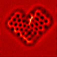
Same background-substracted image as above but with a red color scale to highlight the heart-shape structure of the molecule.