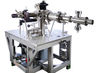
Photoemission Electron Microscopy (PEEM) is an extremely powerful imaging technique, whose versatility for topographical, chemical and magnetic contrast imaging at high resolution has been demonstrated in many laboratory and synchrotron applications.
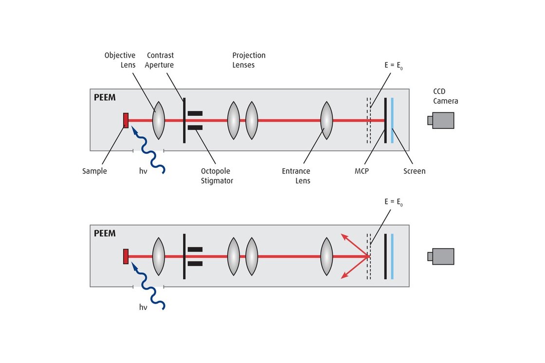
Important contributions to the characterization of magnetic devices, Plasmon research, surface chemistry and high lateral resolution chemical analysis in combination with synchrotron radiation, investigation of time resolved processes and k-space imaging are only a few examples of active PEEM based research. In contrast to a Scanning Electron Microscope (SEM), PEEM directly images surface areas emitting photoelectrons in real-time, without scanning.
Electron emission from surfaces can be caused in various ways - by photon irradiation excitation, thermally, via electron/ion bombardment or by field emission. Over the years the FOCUS-PEEM has been continually improved in performance and usability. The PEEM together with the dedicated high stability integrated sample stage (IS) and various available energy filters follow a modular concept easy to upgrade. In addition software assisted operation ensures time efficient and secure PEEM operation even on challenging samples. With more than 50 units in the market the FOCUS IS-PEEM is a powerful surface analysis tool.
More Information
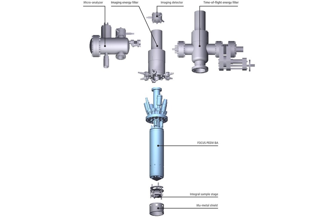
The Modular Concept
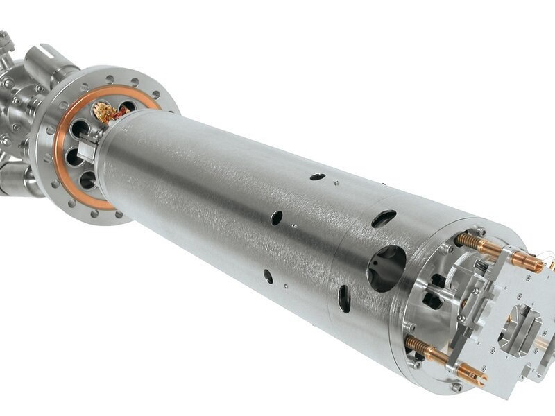
The FOCUS PEEM BA is the basic version of the PEEM instrument series. It is a bolt-on instrument which can be easily integrated in vacuum chambers. It is the heart of all extended PEEM instruments. The modular design of the PEEM allows adapting the instrument to the individual needs of the planned applications.
The integral sample stage of the IS-PEEM guarantees ultimate stability, various energy filters allow for optimum spectroscopy conditions, the position read out for the sample and aperture positioning offers e.g. fast re-allocation of small sample features, a dedicated LHe cooled high precision 4 axis sample stage allows sample analysis at < 30 K together with eucentric sample rotation a k-space (transfer) lens allows for local ARUPS analysis with exceptional large angle acceptance of ± 1.8 Å-1.
Imaging Energy Filter (IEF)
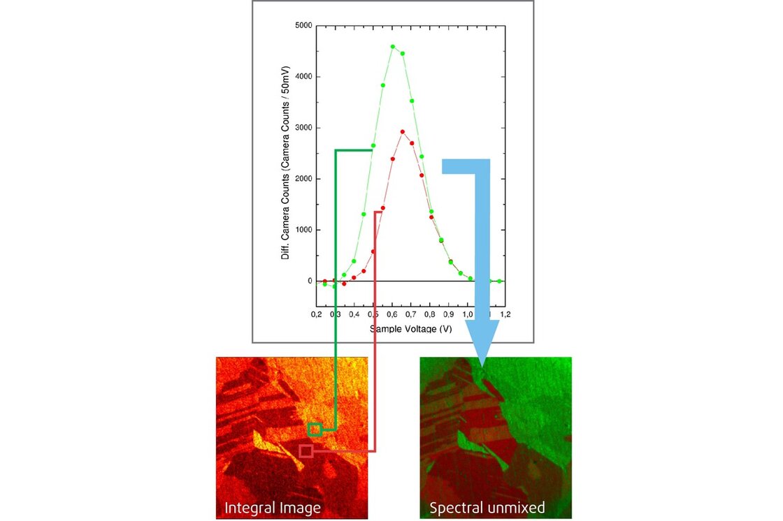
The retarding imaging energy filter (IEF) acts as a high pass filter for the full image. Setting the energy filter to a specific electron energy facilitates element mapping with increased contrast.
The IEF is a dedicated energy filter for all applications with good photoelectron yield. The IEF effectively reduces the chromatic aberration, i.e. the energy spread of the electrons contributing to the formation of the image. This is of particular importance for chemical and magnetic imaging in synchrotron applications, where the achievable resolution is reduced due to the “unfocused” contribution of high energy electrons.
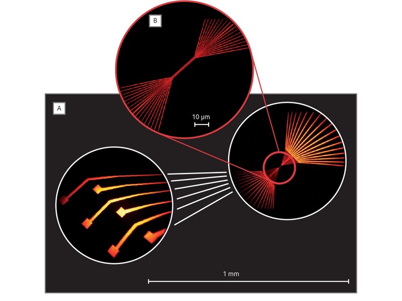
The IEF can also be used for micro spectroscopy analysis. XPS and AES spectra up to a kinetic energy of 1600 eV and with energy resolution well below 100 meV can be obtained in areas smaller than 1 μm.
Time Of Flight Energy Filter
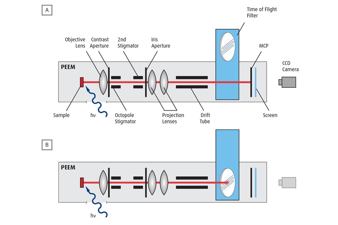
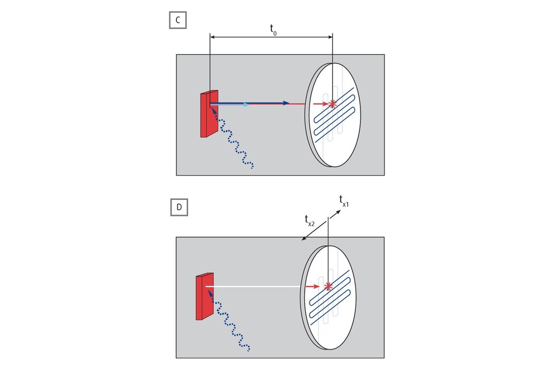
The Time Of Flight (TOF) imaging energy filter is ideally suited for energy filtered low intensity applications with electrons in the low kinetic energy range. Together with a time sensitive imaging Delay Line Detector (2D DLD), a dedicated drift tube and a pulsed light source, the TOF PEEM offers a unique detection system.
The detector allows true single electron counting with massive parallel detection and excellent signal to noise ratio. In addition to standard energy filtered TOF PEEM experiments the detector also allows for time dependent studies with an overall time resolution of < 250 ps.
Lower drift energies inside the drift tube allow higher energy resolution. The special design of the FOCUS PEEM allows for operation at extremely low drift energies (10 eV) while maintaining the lateral resolution of the instrument. The TOF PEEM has achieved an energy resolution of down to 50 meV. A prerequisite for any kind of TOF experiment is a pulsed light source such as pulsed lasers or synchrotrons.
The TOF detector may be retrofitted to existing FOCUS (IS-)PEEMs.
Specifications
50 .. 16000
30, 70, 150, 500, 1750
optionally
3 .. 800
< 40
optional, see below
MCP + CCD camera
single or double
up to 60 sec/ frame, 25 frames/sec
up to 900 sec/frame, 50 frames/sec
up to 10 sec/frame, 140 frames/sec
optional, double MCP needed
ProNanoESCA
optional
Energy filter: retarding field
Energy range [eV]: 0 .. 1595
Achievable energy resolution [meV]: < 50
Energy filter: drift tube with DLD detector
Energy range [eV]: 0 .. 1595
Achievable energy resolution [meV]: < 30
Extended Real space field of view [µm]: 2.5 .. 1700
k-space Field of View [Å-1]: > 6
Achievable k-space resolution [Å-1]: 0.018
For full specifications and more information about product options, please do not hesitate to contact your local sales representative. Find the contact details here: Contact Us
Reference Systems
Downloads
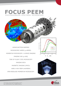
FOCUS PEEM: Photo Emission Electron Microscope
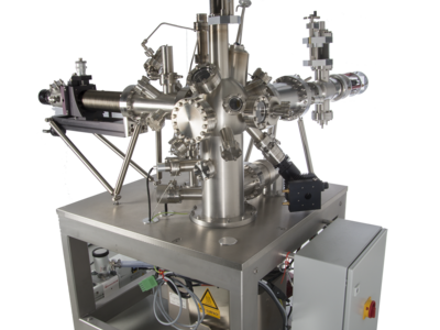
The FOCUS PEEM utilises the technique of photoemission electron microscopy to image electrons emitted from any flat and conducting sample surface. Today the FOCUS PEEM is designed to combine both real- and k-space imaging together with energy filtering and hence spectroscopic microscopy (“spectromicroscopy”) at its best
