Altermagnetic Band Splitting in 10 nm Epitaxial CrSb Thin Films
Result of the Month
Author: Sandra Santhosh, Paul Corbae, Wilson J. Yánez-Parreño, Supriya Ghosh, Christopher J. Jensen, Alexei V. Fedorov, Makoto Hashimoto, Donghui Lu, Julie A. Borchers, Alexander J. Grutter, Timothy R. Charlton, Saurav Islam, Diana Golovanova, Yufei Zhao, Aria Tauraso, Anthony Richardella, Binghai Yan, K. Andre Mkhoyan, Christopher J. Palmstrøm, Yongxi Ou, Nitin Samarth
Institute:
''Samarth Group, Dept. of Physics, Pennsylvania State University, USA''
Advanced Materials
URL: https://doi.org/10.1002/adma.202508977
Date: 7/2026
Instruments:
ARPES Lab, EVO 25/50 MBE, DA30-L
Altermagnets are a newly identified family of collinear antiferromagnets with a momentum-dependent spin-split band structure of non-relativistic origin, derived from spin-group symmetry-protected crystal structures. Among candidate altermagnets, CrSb is attractive for potential applications because of a large spin-splitting near the Fermi level and a high Néel transition temperature of around 700 K. Molecular beam epitaxy is used to synthesize CrSb (0001) thin films with thicknesses ranging from 10 to 100 nm. Structural characterization, using reflection high energy electron diffraction, scanning transmission electron microscopy, and X-ray diffraction, demonstrates the growth of epitaxial films with good crystallinity. Polarized neutron reflectometry shows the absence of any net magnetization, consistent with antiferromagnetic order. In vacuo angle resolved photoemission spectroscopy (ARPES) measurements probe the band structure in a previously unexplored regime of film thickness, down to 10 nm. These ARPES measurements show a bulk-type, 3D momentum-dependent band splitting of up to 0.7 eV with g-wave symmetry, consistent with that seen in prior studies of bulk single crystals. The distinct altermagnetic band structure required for potential spin-transport applications survives down to the ∼10 nm thin film limit at room temperature.
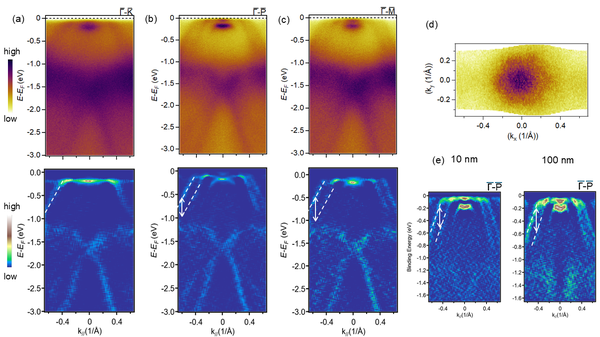
Laboratory-based He lamp ARPES measurements of CrSb thin films. a–c) ARPES spectra of CrSb films (10 nm thickness) along different in-plane momenta direction (top) and corresponding 2D curvature images (bottom).
The color lines are guides to the eyes for the split bands in CrSb. Fermi surface map d) of CrSb (10 nm thickness), e) comparison of the split bands in CrSb films of 10 nm and 100 nm thickness.
Test measurements to optimise growth condition and thickness dependence were done at laboratory-based in vacuo ARPES Lab connected to EVO-50 MBE, located at Pennsylvania State University
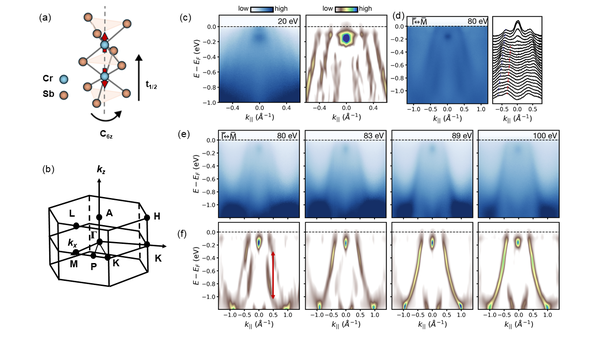
Synchrotron-based Photon energy dependent ARPES measurements of 10 nm CrSb thin films. a) Schematics of the crystal symmetry and b) the Brillouin zone of CrSb. c) cut at hν = 20 eV and corresponding 2D curvature plot showing evident band splitting. d) cut at hν = 80 eV and corresponding MDC showing evident band splitting superimposed on a relatively large background.
Blue and red lines are guide to the eye. e) cuts from hν = 80 eV to 100 eV. Band splitting disappears closer to hν = 100 eV which corresponds to the bulk A point. f) 2D curvature plots of the spectra shown in (e) to emphasize splitting. The vertical arrow shows a maximum splitting of 700 meV in the hν = 80 eV data.
MBE grown samples at Pennsylvania State University were transferred to synchrotrons using UHV suitcase.
3D band structure was measured using synchrotron-based ARPES, BL 5-2 at Stanford Synchrotron Radiation Lightsource and BL 10.0.1.2 at Advanced Light Source.