

Impetus MIM
Momentum microscope with hemispherical energy analyser and multi-mode front lens
- Energy-resolved real-space and k-space microscopy with imaging hemispherical energy analyser
- Extension for sub-µm ARPES, XPEEM and dark-field imaging using enlarged aperture arrays
- Multi-mode front lens for adaptable extraction-field conditions
- Flexible UV, VUV, laser or synchrotron excitation integration
Impetus MIM is Scienta Omicron’s advanced Momentum and Imaging Microscopy platform for energy-resolved real-space and k-space photoemission microscopy. It combines a momentum and imaging microscope with a single hemispherical energy analyser, microchannel plate/camera detection, and a multi-mode front lens (German patent DE10 2017 126 882 B3).
Developed in a joint effort with Momentum Microscopy pioneer Professor Gerd Schönhense (University of Mainz), Impetus MIM brings together new microscope concepts with Scienta Omicron’s experience in high-resolution ARPES and complex UHV systems.
Advanced zoom optics, piezo-driven contrast and field apertures, and a double mu-metal-shielded lens column support controlled navigation between real-space features and k-space information. The extension for sub-µm ARPES, XPEEM and dark-field imaging (German patent DE10 2020 104 151 B3) expands the workflows available from selected regions of interest by including enlarged aperture arrays for field and contrast apertures.
The hemispherical energy analyser uses a 200 mm mean radius and supports energy selection over defined pass-energy settings. Detection is based on a microchannel plate, phosphor screen and camera, with simple switching between image mode and event mode for integrated-intensity imaging or single-event counting at low intensity.
The chamber can be configured with a UV LED for alignment, a small-spot VUV gas discharge lamp, or a port for synchrotron or laser connection. A multitude of upgrades and system extensions is available to adapt the Impetus MIM Lab as your research needs evolve.
More Information
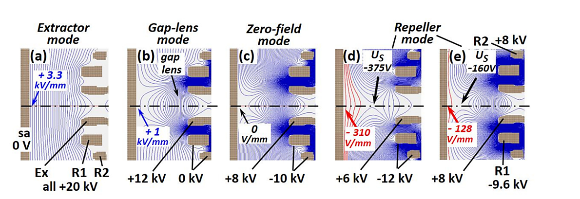
Fig. 1. Operating schemes for the various modes of the new front lens, with the contours of equipotential surfaces. (a) Classic extractor mode with the extractor Ex and ring electrodes R1 and R2 at 20 kV, resulting in a homogeneous field of F=+3.3 kV/mm. (b) Gap-lens mode (thin black line), UEx=12 kV and UR1=UR2=0, resulting in the formation of an additional lens in front of Ex, which reduces the field to F=+1 kV/mm at the sample. (c) Zero-field mode (F=0), achieved with UEx=8 kV and negative ring electrodes (UR1=UR2=-10 kV). (d) Repeller mode, here with F=-310 V/mm, attained with UEx=6 kV and UR1=UR2=-12 kV. The saddle point at US=-375 V defines the low-energy cut-off. (e) Repeller mode, modified by setting UEx=UR2=+8 kV and UR1=-9.6 kV. In (d,e) the retarding field is indicated by red potential contours.Illustration of various multi-mode lens settings (image and caption from https://arxiv.org/pdf/2408.10104)
Further details can be found in the following publications:
(1) O. Tkach, G. Schönhense: “Multimode objective lens for momentum microscopy and XPEEM: Theory”, Ultramicroscopy 276 (2025) 114167
(2) O. Tkach, S. Fragkos, D. Biswas, J. Liu, O. Fedchenko, Y. Lytvynenko, S. Babenkov, D. Zimmer, Q. L. Nguyen, S. Chernov, D. Kutnyakhov, M. Scholz, N. Wind, A. Gloskovskii, F. Pressacco, J. Dilling, L. Bruckmeier, M. Heber, L. Wenthaus, G. Brenner, D. Puntel, P. E. Majchrzak, D. Liu, F. Scholz, J. A. Sobota, J. D. Koralek, G. Dakovski, A. Mehta, N. Sirica, M. Hoesch, C. Schlueter, L. V. Odnodvorets, Y. Mairesse, T.-L. Lee, A. Kunin, K. Rossnagel, Z. X. Shen, H.-J. Elmers, S. Beaulieu, G. Schönhense: “Multimode objective lens for momentum microscopy and X-ray photoemission electron microscopy: Experiments”, Rev. Sci. Instrum. 97, 033703 (2026)
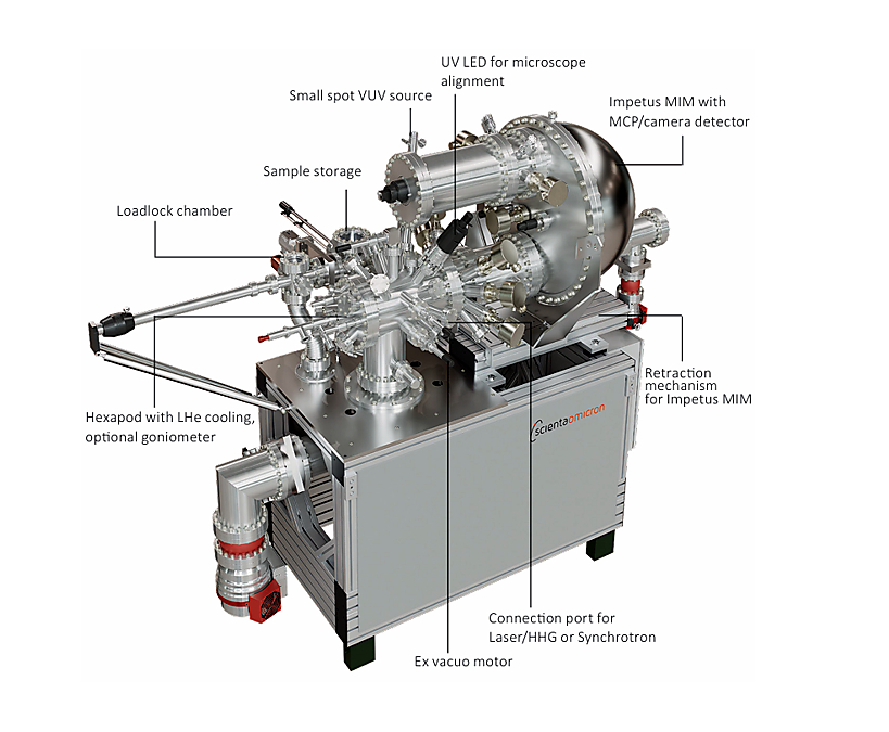
Momentum microscope with hemispherical energy analyser and multi-mode front lens
- Lens column consisting of
- Special objective lens
- Two sets of zoom optics to create intermediate real space and k-space images, each zoom optic with octopole stigmator and deflector
- Arrays of 7 contrast and 16 field apertures with UHV compatible, non-magnetic piezo motors with encoders
- Double mu-metal shield enclosing the lens elements
- Extension for sub-µm ARPES, XPEEM, and dark-field imaging (German patent DE10 2020 104 151 B3), including enlarged aperture arrays for field and contrast apertures
- Multi-mode front lens for reduction of the extraction field at the sample or suppression of space charge effects due to slow electrons (German patent DE10 2017 126 882 B3)
- Hemispherical energy analyser (HEA) with 200 mm mean radius
- Maximum acceptance angle: ± 90°
- Kinetic energy range: 0-2000 eV
- HEA pass energy range 2-200 eV with pre-defined settings in the range 2-25 eV and further settings for higher pass energies
- Detector unit using a microchannel plate plus phosphor screen and camera
- Simple switching between “image” and “event” detection mode measuring integrated intensity or counting single events at low intensity, respectively
- Data acquisition PC with pre-installed software based on Scienta Omicron’s PEAK
- Energy resolution < 10 meV
- Spatial resolution in PEEM mode < 50 nm
- Minimum region-of-interest in k-mode < 1 µm diameter
- k-space resolution < 0.01 Å-1
High-precision hexapod sample stage with LHe cooling
- ±10 mm x/y travel (lateral) and 25 mm z travel (sample retraction for letting the exciting photon beam pass through), as well as 2 orthogonal tilt motions (±5°) for fine tuning of the sample orientation
- Optional goniometer for 0-200° azimuthal sample rotation
- Ex vacuo motors with encoders
- Manipulator x/y/z precision: 1 µm
- Manipulator rotation precision: 0.5°
- Electrically insulated sample acceptor stage made from Au-coated OFHC copper. Suitable for standard Scienta Omicron sample flags, no special sample plates needed!
- LHe flow cryostat, temperature range: < 15…500 K
Excitation Sources
- UV-LED for pre-alignment without the need for water cooling
- Small spot gas discharge lamp for continuous excitation with VUV light
- 3-stage differential pumping system to maintain minimal background pressure in the microscopy chamber
- Multilayer focusing mirrors
- Port on microscopy chamber for connection to a Synchrotron or laser system
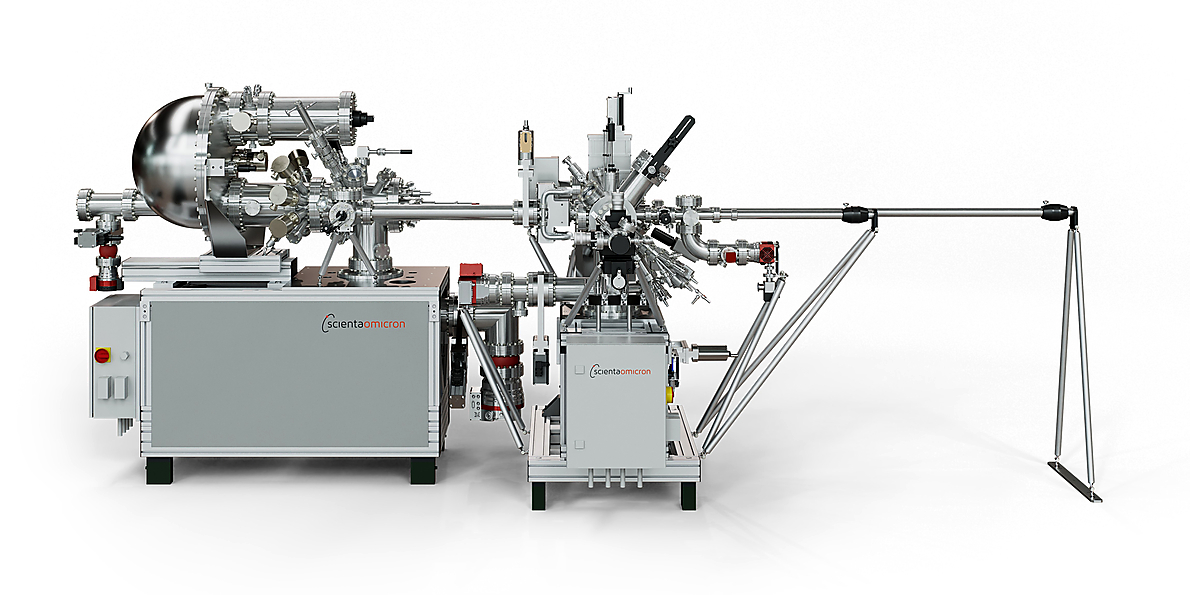
UHV System
- Mu-metal analysis chamber (two layers) with ports for excitation sources, a sample stage, one connection port to a laser or Synchrotron, and several spare ports, e.g. for in vacuum lenses and mirrors or further excitation sources
- Set of UHV pumps (ion getter, TSP, turbo pump, oil-free backing pump) and pressure gauges for the main chamber and the analyser body
- Guaranteed base pressure < 5 x 10⁻¹⁰ mbar
- Scienta Omicron’s MISTRAL control system including comprehensive interlocks for intuitive, user-friendly and fault-proof operation of the UHV system
- Rigid bench for operation of a high-resolution microscope; optional Synchrotron adjustment frame
- Sample storage chamber (10 positions) and separate loadlock for multiple flag-style sample plates, allowing convenient sample handling
- Extension possible, e.g. with Scienta Omicron’s Multiprobe Prep for sample preparation and SPM, or a UHV suitcase for sample exchange with remote UHV systems
Specifications
Momentum and imaging microscope with hemispherical energy analyser and multi-mode front lens
Hemispherical energy analyser, 200 mm mean radius.
Kinetic energy range 0-2000 eV; HEA pass energy range 2-200 eV with pre-defined settings 2-25 eV and further high-pass-energy settings
Energy resolution < 10 meV; PEEM spatial resolution < 50 nm; minimum region-of-interest in k-mode < 1 µm diameter; k-space resolution < 0.01 Å-1
Special objective lens; two zoom-optic sets for intermediate real-space and k-space images, both with octopole stigmator and deflector; double mu-metal shielding
7 contrast and 16 field apertures with UHV-compatible, non-magnetic piezo motors with encoders
Microchannel plate, phosphor screen and camera; image mode and event mode; data acquisition PC with PEAK-based software
High-precision hexapod with LHe cooling; ±10 mm x/y travel; 25 mm z travel; two orthogonal tilt motions ±5°
Optional 0-200° azimuthal goniometer; x/y/z precision 1 µm; rotation precision 0.5°; LHe flow cryostat < 15 K to 500 K
UV LED pre-alignment; small-spot VUV gas discharge lamp with differential pumping and multilayer focusing mirrors; port for synchrotron or laser
Guaranteed base pressure < 5 x 10^-10 mbar; MISTRAL control with interlocks; 10-position sample storage and loadlock; extension possible with Multiprobe Prep for sample preparation and SPM, UHV suitcase exchange, in-vacuum optics/mirrors and additional sources
Impetus MIM is currently available for public pre-launch discussions. For researchers and institutions planning future momentum microscopy investments, Scienta Omicron can support early-stage project planning with budgetary quotations. Please contact us to discuss your requirements.
Downloads
Impetus MIM Brochure
Impetus MIM is Scienta Omicron’s advanced Momentum and Imaging Microscopy platform for energy-resolved real-space and k-space photoemission microscopy. It combines a momentum and imaging microscope with a single hemispherical energy analyser, microchannel plate/camera detection, and a multi-mode front lens.