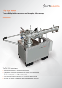
TOF MIM
Time of Flight Momentum and Imaging Microscopy
- Best energy resolution in Momentum Microscopy
- Collection of all electrons emitted from the sample in a 3-dimensional (Ekin ,kx, ky) data cube in a single measurement
- Easy switching between real space and momentum space imaging
- Focus on any feature of interest by piezo-driven adjustable apertures
The global electronic band structure and its dynamics are of great interest in quantum materials. Scienta Omicron’s Time-of-Flight Momentum and Imaging Microscope (ToF MIM) is an efficient tool to characterise global electronic band structure and microstructure at once. It is ideally suited to study ultrafast electron dynamics in pump-probe experiments using ultrashort laser pulses.
More Information
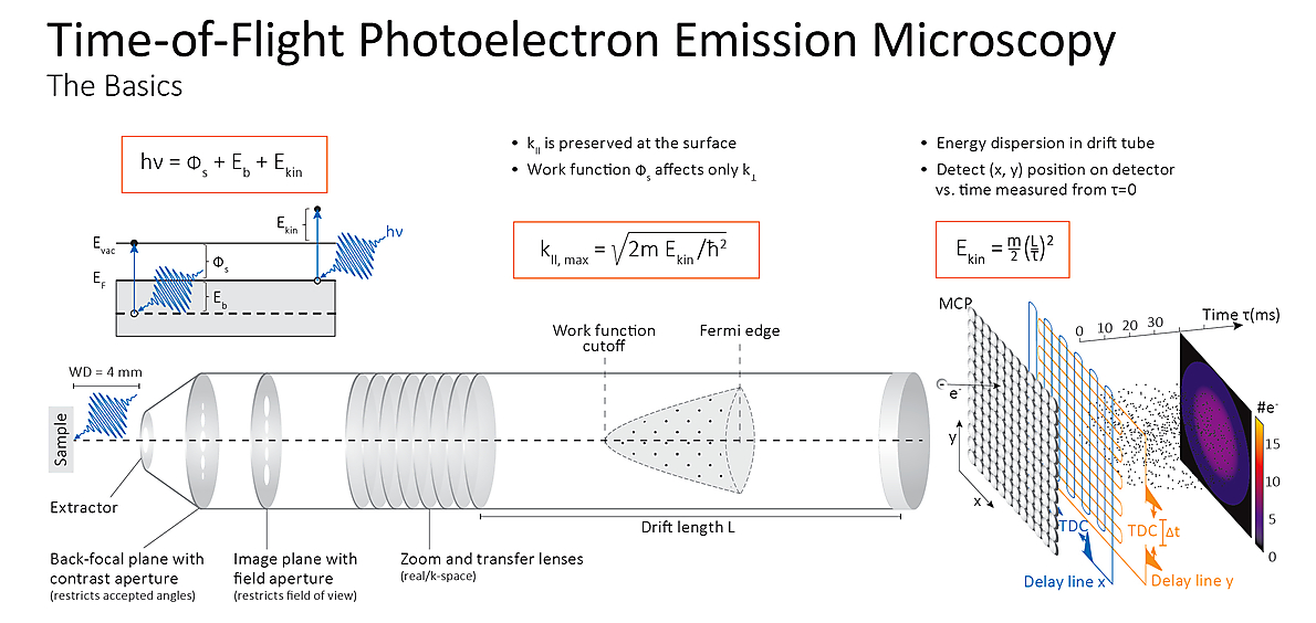
Static Real and Momentum Space Imaging
The ToF Momentum and Imaging Microscope (ToF MIM) captures the full 3D electronic structure in a single measurement, recording either spatial (x, y) or momentum (kx, ky) distributions together with binding energies.
Electrons emitted into the full 2π solid angle are collected in parallel, enabling rapid acquisition of complete datasets without repeating measurements.
Switching between real- and momentum-space imaging is fast and easy, and piezo-driven apertures allow precise control of the field of view without moving the sample.
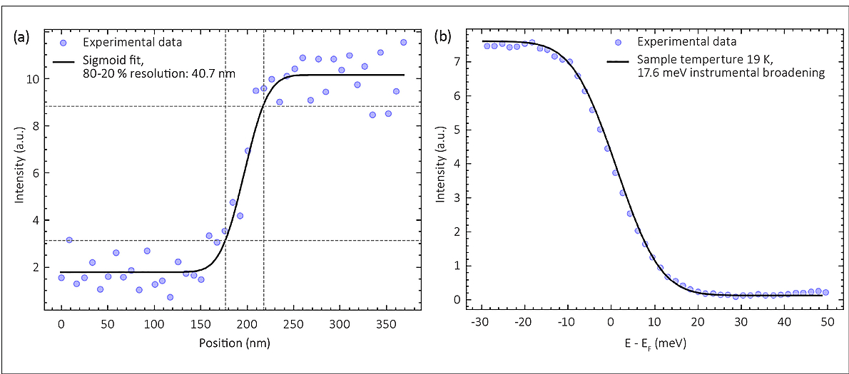
With 40 nm spatial and 17.6 meV energy resolution, ToF MIM is ideal for mapping fine electronic structures on inhomogeneous or nanoscale samples.
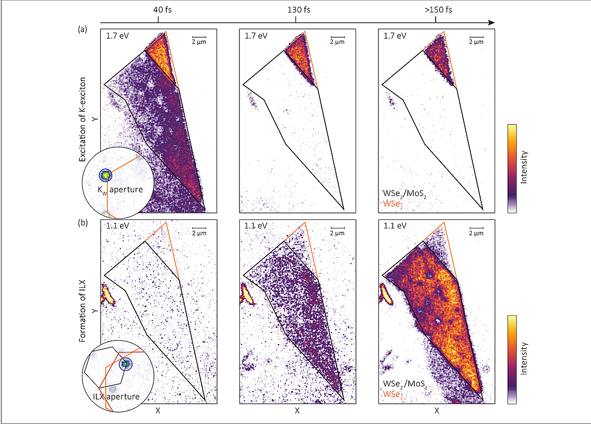
Capturing Ultrafast Dynamics
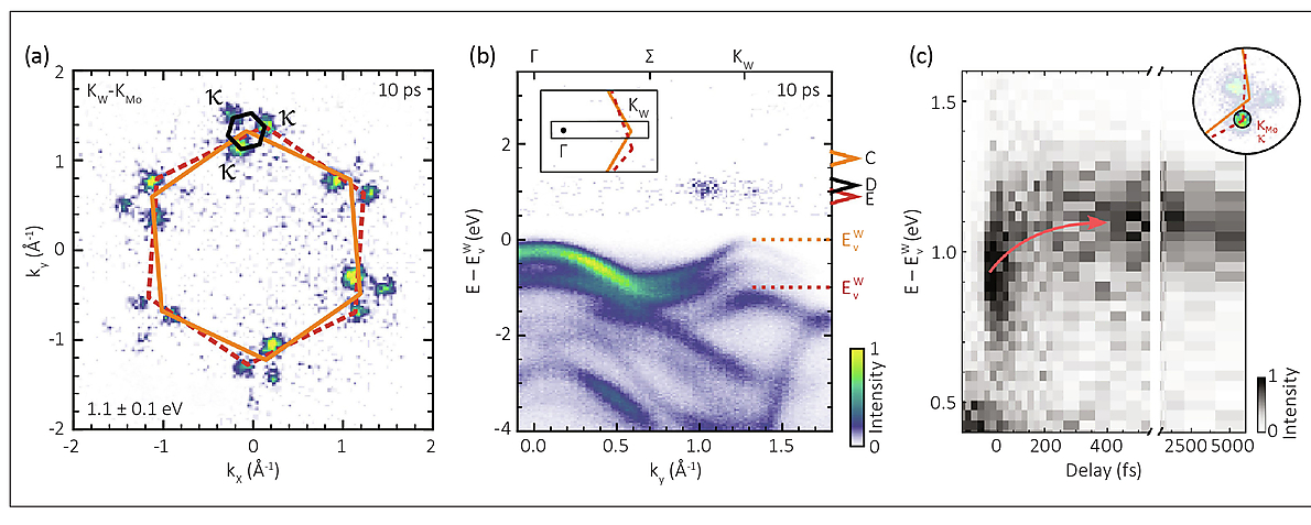
The Time-of-Flight Momentum and Imaging Microscope (ToF MIM) tracks the ultrafast dynamics of hot charge carriers in full energy‑momentum (E‑k) space. Combined with ultrafast EUV/XUV sources like High Harmonic Generation (HHG) or free‑electron lasers, ToF MIM enables 4D data acquisition (energy, momentum, and pump‑probe delay) to reveal global electron dynamics at surfaces.
Its delay‑line detector inherently synchronizes with pulsed light sources, making it ideal for pump–probe experiments. High repetition rates reduce space‑charge effects and support high‑throughput measurements.
A dark-field momentum microscopy mode extends capabilities further, enabling real‑space imaging of selected momenta. This lets researchers directly visualize quasiparticle excitations and their evolution over time, even when optical access is limited.
Ready for Discovery
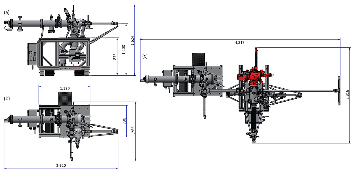
The ToF MIM combines Scienta Omicron’s surface analysis expertise with Surface Concept’s 20 years of delay‑line detector experience, delivering high count rates and reliable performance. The fully motorized hexapod sample stage with LHe flow cryostat ensures precise positioning, long-term stability, and easy maintenance (Figure 6).
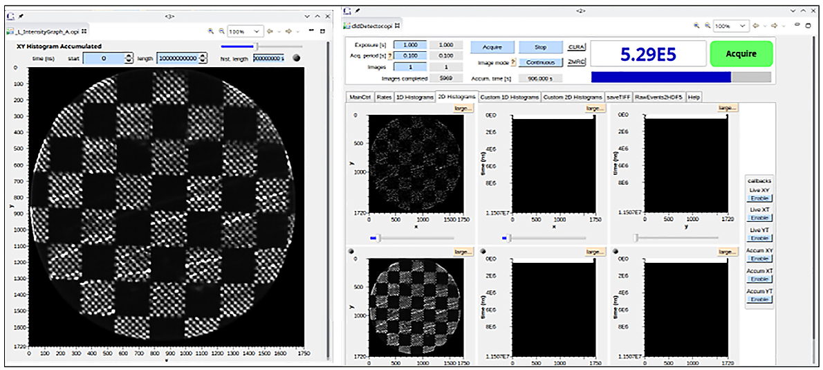
Control and data acquisition use the EPICS framework, enabling intuitive operation, fine-tuning for high-resolution measurements, and flexible data storage in image stacks or open HDF5 event streams (Figure 6).
Many configurable options are available, including connection to the Multiprobe Prep module for rapid sample preparation and multi-technique characterization with evaporators, sputter guns, LEED/Auger, RGA, and VT-XA SPMs (Figure 7).
Specifications
±3 Å-1 (Excitation energy: 40 eV)
±90 degrees
< 0.01 Å-1
< 1 μm (determined by field aperture)
11...1,000 μm
< 50 nm guaranteed
(< 40 nm achieved)
< 25 meV guaranteed
(< 20 meV achieved)
5…500 eV (typically used: 10...30 eV)
Up to 10 eV
250 kHz…2 MHz
80 V...29.9 kV (typically used: 6...20 kV)
Possible using piezo-driven adjustable contrast/field apertures
9 aperture sizes + 200 mesh for alignment
14 aperture sizes + 200 mesh for alignment
4…6 mm (typically used: 4 mm)
5 x 10⁶ cps (assuming a homogeneous distribution)
Hexapod with open cycle LHe cooling
• x, y, z
• Azimuthal rotation
• Tilt around 2 orthogonal axes
All axes are motorised
5 mm x 5 mm x 5 mm
1 μm
±5°
(Optional upgrade: ±90°)
±2°
< 15 K guaranteed
(< 9 K achieved)
400 K (counter heating)
*) All piezo-driven apertures are adjustable in x and y.
DN63CF (horizontal)
Ion getter (300 l/s) and Ti sublimation pump
Optional: turbo-molecular pump (260 l/s)
3x10⁻¹⁰ mbar
UV LED
Electrical sample contacts
Compact mirror integrated into the electron optics allows access under 5° off the sample normal
Parallel spin imaging based on the spin-dependent reflectivity of a Au/Ir target